Biomolecular Models
Objective:
To construct, using models or diagrams, the three dimensional organization of Glucose, Ribose, Maltose, Glycerol, a Triglyceride, a phospholipid, Glycine and Glutamate, at the level of 85% proficiency for each student.
In order to achieve this objective, you will need to be able to:
- Use Bioinformatics to examine the three dimensional structure of biomolecules.
- Use ball and stick models to make biomolecules in three dimension.
- Construct three dimensional models of the simple sugars Glucose and Ribose.
- Explain the effect of changing the orientation of oxygen groups on the biocompatibility of simple sugars.
- Construct a disaccharide, Maltose, from two molecules of Glucose.
- Construct models of Glycerol and the fatty acids linoleic acid and arachidonic acid.
- Construct a Triglyceride from Glycerol and three molecules of a fatty acid.
- Diagram the structure of the phospholipid Phosphatidylcholine
- Construct models of the amino acids Glycine and Glutamate.
- Examine the structure of the 9-amino acid peptide Vasopressin.
Materials:
Group Supplies
Ball and Stick model kit
Computer with the Chime plug-in installed for Internet Explorer
(The Chime plug-in has been pre-installed onto the computers in the physiology lab)
Molecular structure library from the ChemIDplus System at the National Library of Medicine
http://chem.sis.nlm.nih.gov/chemidplus/setupenv.html
(Selected files have been downloaded and pre-loaded onto the computers in the physiology lab)
Methods:
Double click the shortcut named 3d_molecules to select three dimensional reconstructions of biomolecules:

Glucose
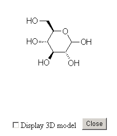
Click on Glucose. A window showing the Fisher projection for Glucose will appear.
Click on the box next to Display 3D model and the three dimensional structure of Glucose will appear.
|
|
|
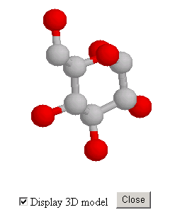
Hold down the left button on the mouse and move the mouse to rotate the molecule.
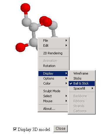
Right click on the molecule to change options for display.
Use the three dimensional image of Glucose to construct a physical model of Glucose using the ball and stick kit. The model on the computer is of Alpha-Glucose. Change the orientation of one Oxygen molecule to make Beta-Glucose.
Maltose
Use the three dimensional image of Maltose to construct a physical model of Maltose using the ball and stick kit. Work with the two groups at your table. Use one model of Alpha-Glucose and one model of Beta-Glucose to make the molecule of Maltose.
Ribose
Use the three dimensional image of Ribose to construct a physical model of Ribose using the ball and stick kit. Make certain that all Oxygen atoms are oriented in the correct direction.
Lipids
Use the three dimensional image of Glycerol to construct a physical model of Glycerol using the ball and stick kit. Examine the structure of the fatty acids Linoleic acid and Arachidonic acid using the three dimensional images.
Begin to construct a Triglyceride using your model of Glycerol and three strands of fatty acids.
Examine the structure of a phospholipid; Phosphatidylcholine, using the three dimensional images.
Amino Acids
Use the three dimensional image of the simple the amino acid Glycine, to construct a physical model of Glycine using the ball and stick kit.
Examine the three dimensional structure of structure of a slightly more complex amino acid, Glutamate. Pay particular attention to the difference between Glycine and Glutamate
Examine the complex structure of the 9-amino acid peptide Vasopressin, using the three dimensional images.
Discussion:
- Compare and contrast the structure of carbohydrates, lipids, and proteins.
- Compare and contrast the dehydration synthesis and hydrolysis of carbohydrates, lipids, and proteins.
- Speculate on whether a carbohydrate, a lipid, or a protein would make a better chemical messenger.
© David G. Ward, Ph.D. Last modified by wardd 23 May, 2006